|
Cell Signaling Technology Inc
c caspase 3 C Caspase 3, supplied by Cell Signaling Technology Inc, used in various techniques. Bioz Stars score: 99/100, based on 1 PubMed citations. ZERO BIAS - scores, article reviews, protocol conditions and more https://www.bioz.com/product/c+caspase+3+affinity/Caspase-3+Antibody/ppr0762015-53-31-34 Average 99 stars, based on 1 article reviews
c caspase 3 - by Bioz Stars,
2026-10
99/100 stars
|
Buy from Supplier |
|
Affinity Biosciences
c caspase3  C Caspase3, supplied by Affinity Biosciences, used in various techniques. Bioz Stars score: 86/100, based on 1 PubMed citations. ZERO BIAS - scores, article reviews, protocol conditions and more https://www.bioz.com/product/c+caspase+3+affinity/3+anti+caspase+cleaved/pmc12857284-58-3-12 Average 86 stars, based on 1 article reviews
c caspase3 - by Bioz Stars,
2026-10
86/100 stars
|
Buy from Supplier |
|
Becton Dickinson
anti-active caspase 3 Anti Active Caspase 3, supplied by Becton Dickinson, used in various techniques. Bioz Stars score: 90/100, based on 1 PubMed citations. ZERO BIAS - scores, article reviews, protocol conditions and more https://www.bioz.com/product/c+caspase+3+affinity/anti+caspase+3/pm16818780-83-27-33 Average 90 stars, based on 1 article reviews
anti-active caspase 3 - by Bioz Stars,
2026-10
90/100 stars
|
Buy from Supplier |
|
Proteintech
anti caspase 3 Anti Caspase 3, supplied by Proteintech, used in various techniques. Bioz Stars score: 97/100, based on 1 PubMed citations. ZERO BIAS - scores, article reviews, protocol conditions and more https://www.bioz.com/product/c+caspase+3+affinity/Caspase+3%2Fp17%2Fp19+Antibody/pm36099983-127-20-50 Average 97 stars, based on 1 article reviews
anti caspase 3 - by Bioz Stars,
2026-10
97/100 stars
|
Buy from Supplier |
|
Cell Signaling Technology Inc
rabbit polyclonal anti caspase 3 Rabbit Polyclonal Anti Caspase 3, supplied by Cell Signaling Technology Inc, used in various techniques. Bioz Stars score: 93/100, based on 1 PubMed citations. ZERO BIAS - scores, article reviews, protocol conditions and more https://www.bioz.com/product/c+caspase+3+affinity/Epitope+Tag+Antibody+Sampler+Kit/pmc03946191-279-13-18 Average 93 stars, based on 1 article reviews
rabbit polyclonal anti caspase 3 - by Bioz Stars,
2026-10
93/100 stars
|
Buy from Supplier |
|
R&D Systems
anti human mouse active caspase 3 antibody Anti Human Mouse Active Caspase 3 Antibody, supplied by R&D Systems, used in various techniques. Bioz Stars score: 92/100, based on 1 PubMed citations. ZERO BIAS - scores, article reviews, protocol conditions and more https://www.bioz.com/product/c+caspase+3+affinity/Human+Caspase-3+Antibody/pmc02743138-103-13-17 Average 92 stars, based on 1 article reviews
anti human mouse active caspase 3 antibody - by Bioz Stars,
2026-10
92/100 stars
|
Buy from Supplier |
|
Boster Bio
antibody against c caspase 3 Antibody Against C Caspase 3, supplied by Boster Bio, used in various techniques. Bioz Stars score: 96/100, based on 1 PubMed citations. ZERO BIAS - scores, article reviews, protocol conditions and more https://www.bioz.com/product/c+caspase+3+affinity/Anti-Caspase-3+CASP3+Antibody/pmc11299798-82-7-27 Average 96 stars, based on 1 article reviews
antibody against c caspase 3 - by Bioz Stars,
2026-10
96/100 stars
|
Buy from Supplier |
|
Wuhan Sanying Biotechnology
c caspase3  C Caspase3, supplied by Wuhan Sanying Biotechnology, used in various techniques. Bioz Stars score: 86/100, based on 1 PubMed citations. ZERO BIAS - scores, article reviews, protocol conditions and more https://www.bioz.com/product/c+caspase+3+affinity/3+anti+caspase/pmc13147163-122-23-27 Average 86 stars, based on 1 article reviews
c caspase3 - by Bioz Stars,
2026-10
86/100 stars
|
Buy from Supplier |
|
Cell Signaling Technology Inc
tbst Tbst, supplied by Cell Signaling Technology Inc, used in various techniques. Bioz Stars score: 97/100, based on 1 PubMed citations. ZERO BIAS - scores, article reviews, protocol conditions and more https://www.bioz.com/product/c+caspase+3+affinity/Caspase-1+Antibody/pmc02722704-91-43-51 Average 97 stars, based on 1 article reviews
tbst - by Bioz Stars,
2026-10
97/100 stars
|
Buy from Supplier |
|
Proteintech
4◦c 4◦C, supplied by Proteintech, used in various techniques. Bioz Stars score: 96/100, based on 1 PubMed citations. ZERO BIAS - scores, article reviews, protocol conditions and more https://www.bioz.com/product/c+caspase+3+affinity/Rac1+Antibody/pm39765117-79-22-25 Average 96 stars, based on 1 article reviews
4◦c - by Bioz Stars,
2026-10
96/100 stars
|
Buy from Supplier |
|
R&D Systems
primary antibody anti caspase 3 Primary Antibody Anti Caspase 3, supplied by R&D Systems, used in various techniques. Bioz Stars score: 96/100, based on 1 PubMed citations. ZERO BIAS - scores, article reviews, protocol conditions and more https://www.bioz.com/product/c+caspase+3+affinity/Human%2FMouse+Active+Caspase-3+Antibody/10__1128_slash_jvi__80__10__4801___4819__2006-150-44-55 Average 96 stars, based on 1 article reviews
primary antibody anti caspase 3 - by Bioz Stars,
2026-10
96/100 stars
|
Buy from Supplier |
|
Cell Signaling Technology Inc
immunocytochemistry rabbit anti cleaved caspase 3 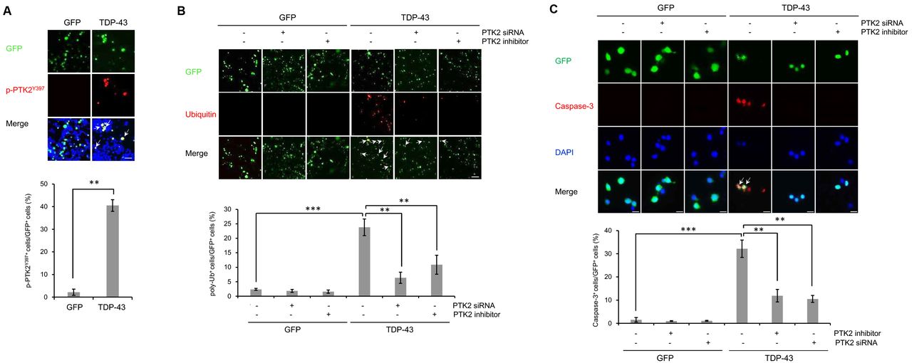 Immunocytochemistry Rabbit Anti Cleaved Caspase 3, supplied by Cell Signaling Technology Inc, used in various techniques. Bioz Stars score: 96/100, based on 1 PubMed citations. ZERO BIAS - scores, article reviews, protocol conditions and more https://www.bioz.com/product/c+caspase+3+affinity/Phospho-(Ser%2FThr)+Akt+Substrate+Antibody/bio_rxiv__355446-33-6-13 Average 96 stars, based on 1 article reviews
immunocytochemistry rabbit anti cleaved caspase 3 - by Bioz Stars,
2026-10
96/100 stars
|
Buy from Supplier |
Image Search Results
Journal: Materials Today Bio
Article Title: A multifunctional and ROS response CO-gas delivery platform for spinal cord regeneration
doi: 10.1016/j.mtbio.2026.102760
Figure Lengend Snippet: COPH could decreases neuronal apoptosis at 7 d.p.i. (A) Immunofluorescence staining of NeuN (green), C-caspase3 (red) and DAPI (blue) in different groups. Scale bar: 50 μm. (B) Western blotting showing the expression of Bax, Bcl-2 and C-caspase3 in different groups 7 d.p.i. (C) Statistical analysis of C-caspase3 fluorescence intensity in each group. (D–F) The quantitative analysis of Bax, Bcl-2 and C-caspase3 protein expression. ∗∗ represents P < 0.01 vs SCI group. ## represent COPs group vs COPH group respectively p < 0.01, (n = 3). (For interpretation of the references to colour in this figure legend, the reader is referred to the Web version of this article.)
Article Snippet: Primary antibody to
Techniques: Immunofluorescence, Staining, Western Blot, Expressing, Fluorescence
Journal: Frontiers in Bioengineering and Biotechnology
Article Title: Uncovering the mechanisms of homologous point acupuncture on knee osteoarthritis through an integrated study of metabolomics and proteomics
doi: 10.3389/fbioe.2026.1791109
Figure Lengend Snippet: Effects of HAP on cartilage autophagy and apoptosis in KOA model rats. Note: (A,C–F) Expression of LC3 I, LC3 II, Beclin1, C-Caspase3 and Bax in cartilage tissue (GAPDH as internal control). (B,G,H) Expression of p62 and Bcl2 in cartilage (GAPDH as internal control). *** p < 0.001, ** p < 0.01, * p < 0.05.
Article Snippet: All the primary antibodies were validated by either the manufacturer or other available publications, listed as follows: HIF1α (Boster, A00013-1), HK2 (Abclonal, A0994),
Techniques: Expressing, Control
Journal: bioRxiv
Article Title: PTK2 regulates the UPS impairment via p62 phosphorylation in TDP-43 proteinopathy
doi: 10.1101/355446
Figure Lengend Snippet: (A-C) N2a cells were transiently transfected with the control vector (pCMV6-AC-GFP) or TDP-43 expression construct (pCMV6-AC-TDP-43-GFP) for 3 days. Cells overexpressing GFP or TDP-43-GFP were treated with ptk2 -specific siRNA (20 nM) or a PTK2 inhibitor (5 μm) for 24 h. Immunocytochemistry was performed thereafter. (A) Immunocytochemistry was subsequently performed to detect PTK2 phosphorylation (p-PTK2 Y397 ; red). Quantification of the percentage of p-PTK2 Y397 -positive cells in GFP-positive cells ( lower ). Arrowheads indicate the co-localization of the p-PTK2 Y397 with TDP-43-GFP-positive cells. Data are presented as the means ± SD of 3 independent experiments. **p<0.005 (Student’s t -test). Scale bars, 200 μm. (B) Immunocytochemistry was subsequently performed to detect ubiquitin (red) or DAPI (nuclei; blue). Quantification of the percentage of poly-ubiquitin-positive staining in GFP-positive cells ( lower ). Arrowheads indicate the co-localization of the poly-ubiquitin with TDP-43-GFP-positive cells. Data are presented as the means ± SD of 3 independent experiments. **p<0.005, ***p<0.001 (two-way ANOVA with Bonferroni multiple comparison test). Scale bars, 20 μm. (C) Immunocytochemistry was subsequently performed to detect the expression of cleaved caspase-3 (red) or DAPI (nuclei; blue). Quantification of the percentage of cleaved caspase-3-positive staining in GFP-positive cells (lower). Arrowheads indicate the co-localization of the caspase-3 with TDP-43-GFP-positive cells. Data are presented as the means ± SD of 3 independent experiments. **p<0.005, ***p<0.001 (two-way ANOVA with Bonferroni multiple comparison test). Scale bars, 200 μm.
Article Snippet: The following antibodies were used for
Techniques: Transfection, Plasmid Preparation, Expressing, Construct, Immunocytochemistry, Staining

Journal: bioRxiv
Article Title: PTK2 regulates the UPS impairment via p62 phosphorylation in TDP-43 proteinopathy
doi: 10.1101/355446
Figure Lengend Snippet: A-B , The p62-, and p62 S403A -overexpressing N2a stable cells lines treated with MG132 (5 μm) for 24 h. Immunocytochemistry was performed thereafter. (A) Immunocytochemistry to detect the Cyto-ID autophagy dye (green) or ubiquitin (red) was subsequently performed. Quantification of the percentage of poly-ubiquitin-positive staining in all cells ( left ), the percentage of CytoID-positive and poly-ubiquitin-positive staining in poly-ubiquitin-positive cells ( middle ), or the percentage of CytoID-positive staining in total cells ( right ). Arrowheads indicate the colocalization of the Cyto-ID autophagy dye with poly-ubiquitin in the p62 stable cells lines. Data are presented as the means ± SD of 3 independent experiments. ***p<0.001, n.s. not significant (Student’s t -test). Scale bars, 10 μm. (B) Immunocytochemistry to detect the DDK (p62; green) or poly-ubiquitin (red) was subsequently performed. Quantification of the percentage of p62-positive puncta in poly-ubiquitin-positive cells (lower). Arrowheads indicate the co-localization of p62 with poly-ubiquitin in the p62 stable cells lines. Data are presented as the means ± SD of 3 independent experiments. *p<0.05 (Student’s t -test). Scale bars, 10 μm. C-D , p62-or p62 S403A -overexpressing N2a stable cells lines were transiently transfected with the control vector (pCMV6-AC-GFP) or TDP-43-expression construct (pCMV6-AC-TDP-43-GFP) for 3 days. Immunostaining was performed thereafter. (C) Immunocytochemistry was subsequently performed to detect poly-ubiquitin (red) or DAPI (nuclei; blue). Arrowheads indicate poly-ubiquitin-positive inclusions. Quantification of the percentage of poly-ubiquitin-positive staining in GFP-positive cells ( right ). Data are presented as the means ± SD of 3 independent experiments. **p<0.005 (two-way ANOVA with Bonferroni multiple comparison test). Scale bars, 10 μm. (D) Immunocytochemistry to detect caspase-3 (red) or DAPI (nuclei; blue) was subsequently performed. Arrowheads indicate caspase-3-positive cells. Quantification of the percentage of caspase-3-positive staining in GFP-positive cells (right). Data are presented as the means ± SD of 3 independent experiments. **p<0.005 (two-way ANOVA with Bonferroni multiple comparison test). Scale bars, 10 μm.
Article Snippet: The following antibodies were used for
Techniques: Immunocytochemistry, Staining, Transfection, Plasmid Preparation, Expressing, Construct, Immunostaining